C-Arm Imaging Equipment
Medical Express Trading is offering C-Arm Imaging equipment. Experience the difference of using a C-arm designed for the specific needs in Orthopedics. It delivers the power to penetrate dense anatomy in lumbar and hip regions in small to large patients. Along with the precision to accentuate boney anatomy for clear spine and ortho images. Also, the performance to get the image you need in less time and with fewer exposures as well.
Below is a list of all our C-Arm Imaging Equipment we offer. To learn more, please go to our contact page by Clicking Here!
 9900 Elite
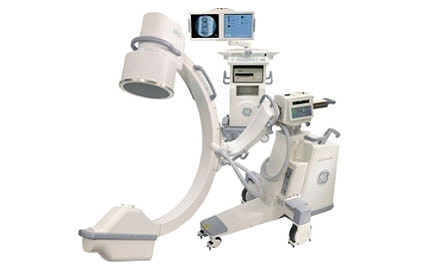
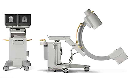
The OEC 9900 Elite C-arm is one of GE Healthcare’s latest c-arm imaging equipment; for sale, available, and approved for use in the United States since August 2012. It was designed to deliver excellent images while maintaining easy-to-use operation, configuration, and C-arm positioning. This is achieved by its intuitive user interface and left-to-right layout. It also has a deeper C arc that allows more space for positioning patients as desired.
The Elite’s image processing power is 100 times greater than older OEC models, utilizing Dynamic Range Management. It is capable of producing 1k x 1k high resolution images. For vascular imaging, it offers the Motion Tolerant Subtraction technique, a feature not previously available on a mobile unit.
The OEC 9900 Elite has a unique battery buffer system, active cooling, and heat monitoring. All these allow for longer procedures. On the other hand, preset imaging profiles shorten procedure set-up times as settings can be changed with a single button.
Available in configurations specific to different fields of practice, you can buy a used GE OEC 9900 Elite C-arm, a truly “elite” model, as a refurbished unit, making it an affordable, cost-effective solution.
OEC 9800 Plus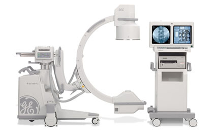
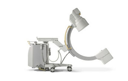
The OEC 9800 C-Arm imaging equipment has led the way for mobile fluoroscopy applications and innovative X-ray imaging technology. It was built on the leadership and experience that comes from having thousands of systems installed worldwide.
Features
- Dual 16″ Square, Flicker Free 1k X 1k High Resolution Monitors
- Tri-Mode 9″/6″/4.5″ Image Intensifier
- Rotating Anode X-ray Tube (0.3mm & 0.6mm) Focal Spots
- 15 kW, 60 Hz High Frequency Generator
- 20 mA Fluoro Boost
- High Resolution 1k x 1k CCD Camera
- 1k x 1k x 16 bit Image Processing
- 63 or 400 Image Storage
- PreView On-Screen Collimator displays allow easy collimator positioning without X-ray exposure
- MANRS (real-time Motion Artifact and Noise Reduction System)
- Digital Zoom Mode. Normal and digital zoom modes provide 2 or 4 times the image size.
- Automatic Digital Brightness and Contrast Controls
- Last Image Hold (LIH)
- Output to Floppy Disk
- Thermal Printer (Optional)
- DICOM (Optional)
- Multiple specialty packages available; GSP, ESP, Vascular, and Neurovascular
OEC 9600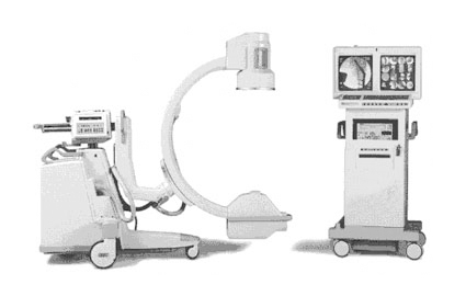
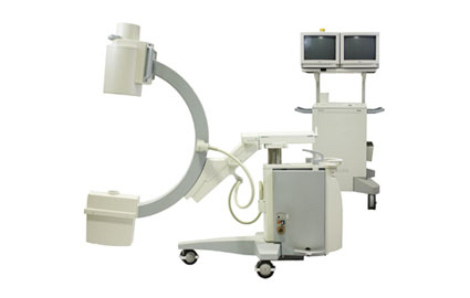
The OEC 9600 C-Arm imaging equipment is a multi-application system that provides solutions to the demanding needs of orthopedic surgery, general surgery, trauma work and pain management.
Features
- Dual 16″ Square High Resolution Flicker Free Monitors
- Tri-Mode 9″/6″/4.5″ Image Intensifier
- Rotating Anode X-ray Tube (0.3mm & 0.6mm) Focal Spots
- 0 kW High Frequency Generator
- 12 mA Fluoro Boost
- High Resolution CCD Camera
- 10 bit Digital Image Processing
- 200 Image Storage
- Collimator with dual opposing shutters
- MARS-Motion Artifact Reduction System
- Frame Averaging
- Last Image Hold (LIH)
- Output to Floppy Disk
- Thermal Printer
- Multiple specialty packages available; SP, GSP, ESP, Vascular and Neurovascular
OEC 9400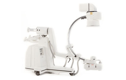
Description for the C-Arm Imaging Equipment
- Digital image storage and review
- Microprocessor control of all functions
- Self-diagnostic program
- User-programmable settings
Specifications
- X-Ray Generator High frequency 2.5 kHz; 7.5 KW full-wave; up to 120 kVp, up to 100 mA for radiographic exposures; fluoro boost/pulsed fluoro capability.
- X-Ray Source Rotating anode; focal spots: 0.3 mm and 1.0 mm; anode heat capacity: 300,000 HU.
- Fluoroscopy Mode Focal spot: 0.3 mm. kVp range: 40-120 kVp. Ma range: 0.2 – 5.0 (20 ma max boost). Continuous one-shot or pulsed fluoro. ABS varies ma, kVp and camera gain. User selects ABS tables for orthopedics, chest, and low dose applications. Collimation is continuously adjustable and rotatable.
- Radiography Mode Focal spot: 0.3 mm and 1.0 mm. kVp range: 50-120 kVp. MAs range: 1 – 300 mAs. Focal spot size automatically selected. Collimation is continuously adjusted and Rotatable.
- Image Intensifier Choice of 6″ or tri-mode 9″/6″/4″ image intensifier. Resolution: -44 lp/cm central (9″ mode), -42 lp/cm peripheral (9″ mode). Contrast ratio: > or equal to 28:1 (9″ mode)
- High Resolution, Low Noise Camera Designed for fluoroscopy. Full 360deg motorized rotation with left-right manipulation. Negative mode. Automatic gain control. Camera tube type: Vidicon. On-screen orientation indicator. Left/right, top/bottom image reversal. Frame averaging (real-time).
- Image Processing Motion artifact reduction (MARS). Digital image enhancement (windowing). Variable edge enhance (track ball). Removable disk (on ESP and above)
- Image Storage Options Radiographic film capability. Sixty-image digital storage. Video tape recorder (VTR). Multi-format camera. New thermal printer.
- Mechanical Free space in arc – 6″ I.I.: 29.5″ (75 cm). Free space in arc: 9″ I.I.: 27.0″ (69 cm). Depth of arc: 23″, movement: 111deg. Travel: 18″ motorized vertically, 8″ horizontally. L-arm rotation: +/-360deg motorized.
- Reversible C-arm: manual (flip-fop).
- Input Power 100/120 VAC, 15 AMP; 200/220/240 VAC, 12 AMP; 50 Hz (optional)
- Main C-arm Frame (stored) and Monitor Cabinet Length: 74.5″ (189 cm)/32″ (81 cm). Height: 66.2″ (168 cm)/65″ (165 cm). Depth: 34″ (86 cm)/27″ (63 cm).
 BV Pulsera
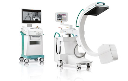
Whether it’s a large field of view for abdominal aortic aneurysm repair, excellent low-dose imaging for interventional pain management or small anatomical detail for cochlear implants, the BV Pulsera’s innovative feature set helps you get the job done for c-arm imaging equipment.
Experience the Difference
- Pulsed acquisition (30 pulses/sec) for better images at a lower dose
- Rotating anode power to penetrate larger patients
- Top quality 3D imaging
- 9″ or 12″ image intensifier
- Excellent maneuverability
Clarity and power
The pulsed exposure mode produces superb image contrast, essentially eliminates movement artifacts in cardiovascular examinations and provides you with virtually blur-free images. In short, the BV Pulsera gives you the power and the image quality your work in the OR demands.
Exceptional heat management capabilities, allow the BV Pulsera to keep pace with your longest cardiovascular and interventional procedures. Rotating anode technology and automatic high penetration mode gives you the power to see through practically any type of patient. See fine details at the steepest projections and observe rapidly moving anatomy.
3D imaging
Optional 3D-RX imaging adds more information about relative position of bony structures, implants and fixation devices. Corrections can be carried out immediately while the patient is still on the table – assisting with better treatment strategy and more streamlined workflow.
In addition, the BV Pulsera with 3D-RX offers you high-quality 3D imaging without compromising full conventional 2D imaging functionality during routine procedures.
Efficiency in the OR
The BV Pulsera’s ultra compact mobile C-arm stand features rear-wheel steering for easy maneuverability and positioning. An optimally designed MobileView Station with ‘touch screen’ monitor display can be moved around the tight confines of a crowded OR, enabling you to get the best viewing position.
With the single user concept and Anatomically Programmed Fluoroscopy (APF) parameters, you can easily control every step of your procedures. This supports high OR throughput.
One system, many applications
The versatility and power of the Philips BV Pulsera mobile surgery system supports many goals. From Bolus Chasing to Abdominal Aortic Aneurysm repair, cardiac procedures to neurosurgical exams, the BV Pulsera has the imaging clarity and power to enable you to do your best work in:
- Cardiovascular procedures
- Orthopedic surgery
- Abdominal surgery
- Neurosurgical procedures
- Thoracic surgery
BV Libra
Designed to handle all your routine procedures, the BV Libra has one of the best price/performance values of any system in its segment. This lightweight, highly maneuverable fluoroscopy system comes with a 9″ image intensifier and loads of important features for c-arm imaging equipment.
Get More for Your Money
- Compact, easy-to-steer
- 9″ triple-mode image intensifier
- BodySmart imaging software
- Dose saving pulsed fluoro mode
- Full RIS/HIS compatibility
Easy to steer, position and operate
Guide the BV Libra right where you need it with hardly any effort. The system’s precision steering and tight turning circle helps you easily maneuver the system through the most crowded spaces.
Once in position, the 9″ image intensifier is ready to handle all your routine procedures including vascular functionality (subtraction and re-masking). Philips optimized imaging chain includes our Charge Coupled Device (CCD) technology and patented anamorphic lens. This means you get exceptional resolution and an outstanding level of detail.
Time saving extras
Acquire excellent image quality with our unique BodySmart software. It immediately finds, tracks and defines the field of view to anatomy – no matter where it is on the image intensifier. Philips sophisticated parallel movement of the system virtually extends your field of view so you can easily image an entire procedure in just one go.
Settings like contrast, brightness and edge enhancements are performed in real-time. All controls are laid out in an intuitive fashion with a minimum of functions and menus.
Reduced X-ray dose
Our real pulsed fluoro mode provides excellent quality images at half the dose of normal fluoroscopy. You get superb images with low X-ray dose for you and your patients.
Efficient data management
Fully integrated DICOM connectivity meets your most demanding requirements. RIS/HIS compatibility allows patient demographics to be imported into the BV Libra system and single images or complete examinations to be exported to your hospital network.
Multi-functional use
The BV Libra is designed to go wherever you need it to – surgery, intensive care, or the ER. It is perfect for such exams and procedures as:
- Orthopedic
- Urological
- Neurosurgical
- Routine vascular exams
- Pain management treatment
- Cholangiograms
BV Endura
Perfect for general fluoroscopy and vascular specialization, the BV Endura helps you clearly view dynamic images in surgery. Designed to improve your workflow, this versatile system has many beneficial characteristics for c-arm imaging equipment.
Features
- Flexible choice between 9″ or 12″ image intensifier
- Extended rotation for vascular projections
- SmartVision imaging technology
- Ultra-compact MobileView Station
It’s about the coverage
Positioned as a mid-level performer, the BV Endura builds on the strengths of the BV Libra by broadening vascular capabilities. Our bigger 12″ image intensifier provides a larger coverage for improved anatomical orientation. And we have extended the C-arm rotation – up to 135o – to provide you with projections necessary for most vascular procedures.
Image enhancement
The next generation in C-arm imaging is SmartVision. This combination of advanced technologies in place across the imaging chain, results in high quality images at a low X-ray dose.
Some Highlights Include
- 1K2 imaging – a fully digital imaging chain with advanced noise reduction and 2D edge enhancements for higher image quality
- BodySmart – software that finds, tracks and defines the field of view to anatomy for ‘first shot right’ imaging
Automatic Shutter Positioning – optimal shutter positioning for the best quality image possible and a clear view of the region of interest - Automatic contrast/brightness – real-time contrast and brightness control at the touch of a button improves ease-of-use and provides faster control for better image quality
Intelligent viewing options
Optimally designed, the MobileView Station’s ultra-compact size makes it easy to move around the tight confines of the OR. And its small footprint ensures it can be brought as close as possible to the operating table.
At the MobileView Station the operator can manually enter patient demographics or retrieve a worklist via the hospital network. Monitors are turned toward the physician at any angle at the start of the procedure and back to the operator at the end to process the images.
- Processed images are immediately sent to PACS
- Two 18″ monitors ensure high quality image viewing
- Monitor height can be quickly adjusted to physician preference
Widely useful
You’ll find the BV Endura can handle a full range of applications including:
- Vascular procedures
- Orthopedic surgery
- Abdominal surgery
- Neurosurgical procedures
- Thoracic surgery
BV 300
- Tri-Mode 9″/6″/4″ Image Intensifier
- 200 Digital Image Storage
- Dual 17″ Hi-resolution Monitors With Cart
- CCD Camera
- Matrix Camera 8″x10″ Format
- Fixed X-ray Tube Certified
- Patient Data Keypad
- BV300 Operators Manuals
- 9″ Tri-Mode Image Intensifier
- 200 Frames Memory
- Hard Copy Device
- Laser Alignment
 Vista Plus
The Ziehm Vista Endo is a unique mobile C-arm imaging equipment system designed in joint co-operation with clinicians to optimise endoscopy procedures under fluoroscopic x-ray control. In order to assist minimally invasive endoscopic procedures (e.g. ERCP/PTCP) the Ziehm Vista Endo’s single mobile cart carries both the fluoroscopic and endoscopic monitors. This configuration allows the endoscopic surgeon the convenience of viewing both the fluoroscopic and color endoscopy image at a single glance. Additionally, the operating team gains more free floor space in the operating room as a separate endoscopic monitor cart becomes unnecessary. The Ziehm Vista Endo is compatible with most popular endoscopes in use today including those from Olympus, Pentax and Fujinon.
Features
- 31 cm image intensifier
- DICOM interfaces to your HIS/RIS/PACS network
- 5 Million Heat Unit Active Cooling x-ray generator for extended operating time
- Digital cine fluoroscopy replay
- 5″ floppy disk or CD-RW for image export and archiving
- Accommodation of video printer as well as video recorder
Vision
Standard Features for this C-Arm Imaging Equipment
- 4/6/9” Tri Mode Image Intensifier
- Dual 18″ Hi Resolution Flat Screen Monitors
- 1k x 1k Resolution
- 45 Degrees Forward Rotation
- Rotating Anode X-Ray Tube
- Touch Screen Controls
- Active Cooling
- DICOM 3.0
- USB Storage
- Vision Center Software
- LIH – Last Image Hold
- Boost Function
- Sharpen Feature
- Artifact Reduction
- High Frequency Generator
- Full Electrical Inspection By Ziehm Technician
- Set To Factory Ziehm Specs
- New Paint
- Sony Paper Printer
- Patient Annotation Keyboard
Options
- DVD Burner
- Flat Image Intensifier
Quantum

Working with the Ziehm Quantum provides fast and cost effective results in a wide variety of medical procedures. The Ziehm Quantum provides high image quality, functionality, mobility, and network connectivity to keep pace with clinical requirements of today’s modern medical practice.
Features
- Most Comprehensive Feature Rich, Full Size C-Arm In a Compact Design
- 1K2 x 16 Bit Highline Video Image Display, Allowing Superb Image Quality
- Simplified, Centralized User Interface
- Integrated 18.1” High Resolution TFT Monitors
- Eliminates Need for Monitor Cart, Providing Excellent Space Saving Maneuverability
- Connectivity Provides DICOM 3.0, USB, DVD
- 4/6/9 Tri-Mode Image Intensifier
- Standard C
- Dual Hi Resolution Monitors On C-Arm
- Digital CCD Camera
- Stationary Anode X-Ray Tube
- Small Footprint
- 45° Forward Orbital Rotation
- ESP Software Package
- Medicapture or Sony Paper Printer